Shoulder Procedures
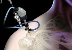
Shoulder Arthroscopy
Arthroscopy is a minimally invasive diagnostic and surgical procedure performed for joint problems. Shoulder arthroscopy is performed using a pencil-sized instrument called an arthroscope. The arthroscope consists of a light system and camera that projects images of the surgical site onto a computer screen for your surgeon to clearly view.
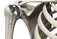
Shoulder Joint Replacement
Total shoulder replacement surgery is performed to relieve symptoms of severe shoulder pain and disability due to arthritis. In this surgery, the damaged articulating parts of the shoulder joint are removed and replaced with artificial prostheses. Replacement of both the humeral head and the socket is called a total shoulder replacement.
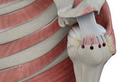
Rotator Cuff Repair
Rotator cuff repair is a surgery to repair an injured or torn rotator cuff. It is usually performed arthroscopically on an outpatient basis. An arthroscope, a small, fiber-optic instrument consisting of a lens, light source, and video camera. The camera projects images of the inside of the joint onto a large monitor, allowing your doctor to look for any damage, assess the type of injury and repair it. Large rotator cuff tears may require open surgery.
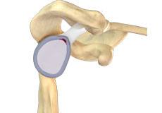
SLAP Repair
A SLAP repair is an arthroscopic shoulder procedure to treat a specific type of injury to the labrum called a SLAP tear.
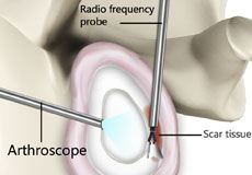
Capsular Release
A capsular release of the shoulder is surgery performed to release a tight and stiff shoulder capsule, a condition called frozen shoulder or adhesive capsulitis. The procedure is usually performed arthroscopically through keyhole-size incisions.
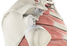
Shoulder Labrum Reconstruction
The shoulder joint is a ball and socket joint. A ball at the top of the upper arm bone (the humerus) fits neatly into a socket, called the glenoid, which is part of the shoulder blade (scapula). The labrum is a ring of fibrous cartilage surrounding the glenoid, which helps in stabilizing the shoulder joint.
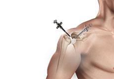
Shoulder Reconstruction Surgery
Shoulder reconstruction surgery is an operative procedure in which stretched or torn soft-tissue structures that surround the shoulder joint such as the capsule, ligaments, and cartilage, are repaired to secure the shoulder joint in place. The procedure is mainly employed for the treatment of individuals with shoulder instability, to prevent recurrent joint dislocations, and to restore normal shoulder range of motion and function.
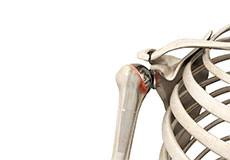
Revision Shoulder Replacement
Revision surgery is usually performed under general anesthesia. You are positioned in such a way as to allow all possible variations in the treatment plan. Incisions are made to gain optimal access to the problem and usually follow previous incisions with extensions made as necessary.
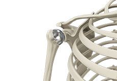
Shoulder Resurfacing
The shoulder is an active joint is prone to injuries and may also get affected by conditions such as arthritis, which results in impaired functioning and related discomfort. The traditional method of treatment for such conditions is shoulder joint replacement. However, advances in technology have resulted in a superior alternative technique known as shoulder resurfacing.
Shoulder Conditions
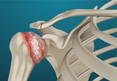
Arthritis of the Shoulder
The term arthritis literally means inflammation of a joint but is generally used to describe any condition in which there is damage to the cartilage. Damage of the cartilage in the shoulder joint causes shoulder arthritis. Inflammation is the body's natural response to injury. The warning signs that inflammation presents are redness, swelling, heat, and pain.
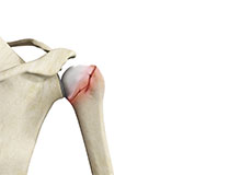
Shoulder Fractures
A break in a bone that makes up the shoulder joint is called a shoulder fracture. The clavicle and end of the humerus closest to the shoulder are the bones that usually get fractured. The scapula, on the other hand, is not easily fractured because of its protective cover by the surrounding muscles and chest tissue.
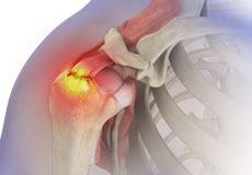
Rotator Cuff Tear
A rotator cuff is a group of tendons in the shoulder joint that provides support and enables a wide range of motion. A major injury to these tendons may result in rotator cuff tears. It is one of the most common causes of shoulder pain in middle-aged and older individuals.
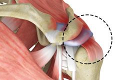
Rotator Cuff Pain
The rotator cuff consists of a group of tendons and muscles that surround and stabilize the shoulder joint. These tendons allow a wide range of movement of the shoulder joint across multiple planes. Irritation or injury to these tendons can result in rotator cuff pain.
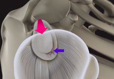
Shoulder Labral Tear
Traumatic injury to the shoulder or overuse of the shoulder (throwing, weightlifting) may cause the labrum to tear. In addition, aging may weaken the labrum leading to injury.
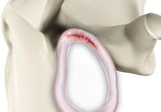
SLAP Tears
The term SLAP (superior –labrum anterior-posterior) lesion or SLAP tear refers to an injury of the superior labrum of the shoulder. The most common causes include falling on an outstretched arm, repetitive overhead actions such as throwing and lifting a heavy object. Overhead and contact sports may put you at a greater risk of developing SLAP tears.
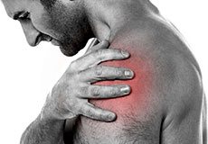
Shoulder Pain
Pain in the shoulder may suggest an injury, which is more common in athletes participating in sports such as swimming, tennis, pitching, and weightlifting. The injuries are caused due to the over usage or repetitive motion of the arms.
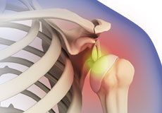
Shoulder Dislocation
Sports that involve overhead movements and repeated use of the shoulder at your workplace may lead to sliding of the upper arm bone from the glenoid. The dislocation might be a partial dislocation (subluxation) or a complete dislocation causing pain and shoulder joint instability. The shoulder joint often dislocates in the forward direction (anterior instability), and sometimes in the backward or downward direction.
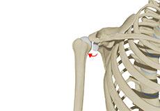
Posterior Shoulder Instability
Posterior shoulder instability, also known as posterior glenohumeral instability, is a condition in which the head of the humerus (upper arm bone) dislocates or subluxes posteriorly from the glenoid (socket portion of the shoulder) as a result of significant trauma. A partial dislocation is referred to as a subluxation whereas a complete separation is referred to as a dislocation.
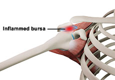
Rotator Cuff Bursitis
The rotator cuff is a set of muscles and tendons which hold the various bones of the shoulder joint together, providing strength and support. Inflammation of the bursa, a fluid-filled sac between the rotator cuff tendons and a bony process at the top of the shoulder called the acromion, is known as shoulder bursitis or rotator cuff bursitis. It is often accompanied by inflammation of the tendons or rotator cuff tendonitis.
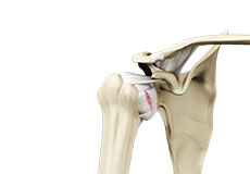
Shoulder Ligament Injuries
Shoulder ligament injuries are injuries to the tough elastic tissues present around the shoulder that connect bones to each other and stabilize the joint. The ligaments present in the shoulder are connected to the ends of the scapula, humerus, and clavicle bones which form the shoulder complex. The extensive stretching or tearing of these ligaments from acute or chronic injuries can lead to instability in the shoulder joint.
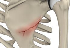
Fracture of the Shoulder Blade
The scapula (shoulder blade) is a flat, triangular bone providing attachment to the muscles of the back, neck, chest and arm. The scapula has a body, neck and spine portion.
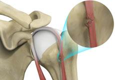
Bicep Tendon Rupture
The biceps muscle is located in the front side of your upper arm and functions to help you bend and rotate your arm. The biceps tendon is a tough band of connective fibrous tissue that attaches your biceps muscle to the bones in your shoulder on one side and the elbow on the other side.
Shoulder Anatomy

The shoulder is the most flexible joint in the body that enables a wide range of movements including forward flexion, abduction, adduction, external rotation, internal rotation, and 360-degree circumduction. Thus, the shoulder joint is considered the most insecure joint of the body, but the support of ligaments, muscles, and tendons function to provide the required stability.
Bones of the Shoulder
The shoulder joint is a ball and socket joint made up of three bones, namely the humerus, scapula, and clavicle.
Humerus
The end of the humerus or upper arm bone forms the ball of the shoulder joint. An irregular shallow cavity in the scapula called the glenoid cavity forms the socket for the head of the humerus to fit in. The two bones together form the glenohumeral joint, which is the main joint of the shoulder.
Scapula and Clavicle
The scapula is a flat triangular-shaped bone that forms the shoulder blade. It serves as the site of attachment for most of the muscles that provide movement and stability to the joint. The scapula has four bony processes - acromion, spine, coracoid and glenoid cavity. The acromion and coracoid process serve as places for attachment of the ligaments and tendons.
The clavicle bone or collarbone is an S-shaped bone that connects the scapula to the sternum or breastbone. It forms two joints: the acromioclavicular joint, where it articulates with the acromion process of the scapula and the sternoclavicular joint where it articulates with the sternum or breast bone. The clavicle also forms a protective covering for important nerves and blood vessels that pass under it from the spine to the arms.
Soft Tissues of the Shoulder
The ends of all articulating bones are covered by smooth tissue called articular cartilage, which allows the bones to slide over each other without friction, enabling smooth movement. Articular cartilage reduces pressure and acts as a shock absorber during movement of the shoulder bones. Extra stability to the glenohumeral joint is provided by the glenoid labrum, a ring of fibrous cartilage that surrounds the glenoid cavity. The glenoid labrum increases the depth and surface area of the glenoid cavity to provide a more secure fit for the half-spherical head of the humerus.
Ligaments of the Shoulder
Ligaments are thick strands of fibers that connect one bone to another. The ligaments of the shoulder joint include:
- Coracoclavicular ligaments: These ligaments connect the collarbone to the shoulder blade at the coracoid process.
- Acromioclavicular ligament: This connects the collarbone to the shoulder blade at the acromion process.
- Coracoacromial ligament: It connects the acromion process to the coracoid process.
- Glenohumeral ligaments: A group of 3 ligaments that form a capsule around the shoulder joint and connect the head of the arm bone to the glenoid cavity of the shoulder blade. The capsule forms a watertight sac around the joint. Glenohumeral ligaments play a very important role in providing stability to the otherwise unstable shoulder joint by preventing dislocation.
Muscles of the Shoulder
The rotator cuff is the main group of muscles in the shoulder joint and is comprised of 4 muscles. The rotator cuff forms a sleeve around the humeral head and glenoid cavity, providing additional stability to the shoulder joint while enabling a wide range of mobility. The deltoid muscle forms the outer layer of the rotator cuff and is the largest and strongest muscle of the shoulder joint.
Tendons of the Shoulder
Tendons are strong tissues that join muscle to bone allowing the muscle to control the movement of the bone or joint. Two important groups of tendons in the shoulder joint are the biceps tendons and rotator cuff tendons.
Bicep tendons are the two tendons that join the bicep muscle of the upper arm to the shoulder. They are referred to as the long head and short head of the bicep.
Rotator cuff tendons are a group of four tendons that join the head of the humerus to the deeper muscles of the rotator cuff. These tendons provide more stability and mobility to the shoulder joint.
Nerves of the Shoulder
Nerves carry messages from the brain to muscles to direct movement (motor nerves) and send information about different sensations such as touch, temperature, and pain from the muscles back to the brain (sensory nerves). The nerves of the arm pass through the shoulder joint from the neck. These nerves form a bundle at the region of the shoulder called the brachial plexus. The main nerves of the brachial plexus are the musculocutaneous, axillary, radial, ulnar and median nerves.
Blood vessels of the Shoulder
Blood vessels travel along with the nerves to supply blood to the arms. Oxygenated blood is supplied to the shoulder region by the subclavian artery that runs below the collarbone. As it enters the region of the armpit, it is called the axillary artery and further down the arm, it is called the brachial artery.
The main veins carrying de-oxygenated blood back to the heart for purification include:
- Axillary vein: This vein drains into the subclavian vein.
- Cephalic vein: This vein is found in the upper arm and branches at the elbow into the forearm region. It drains into the axillary vein.
- Basilic vein: This vein runs opposite the cephalic vein, near the triceps muscle. It drains into the axillary vein.


